



Myelomalacia in Dogs: Clinical Signs, Diagnosis & Treatment

In Brief
|
What Is Myelomalacia in Dogs?
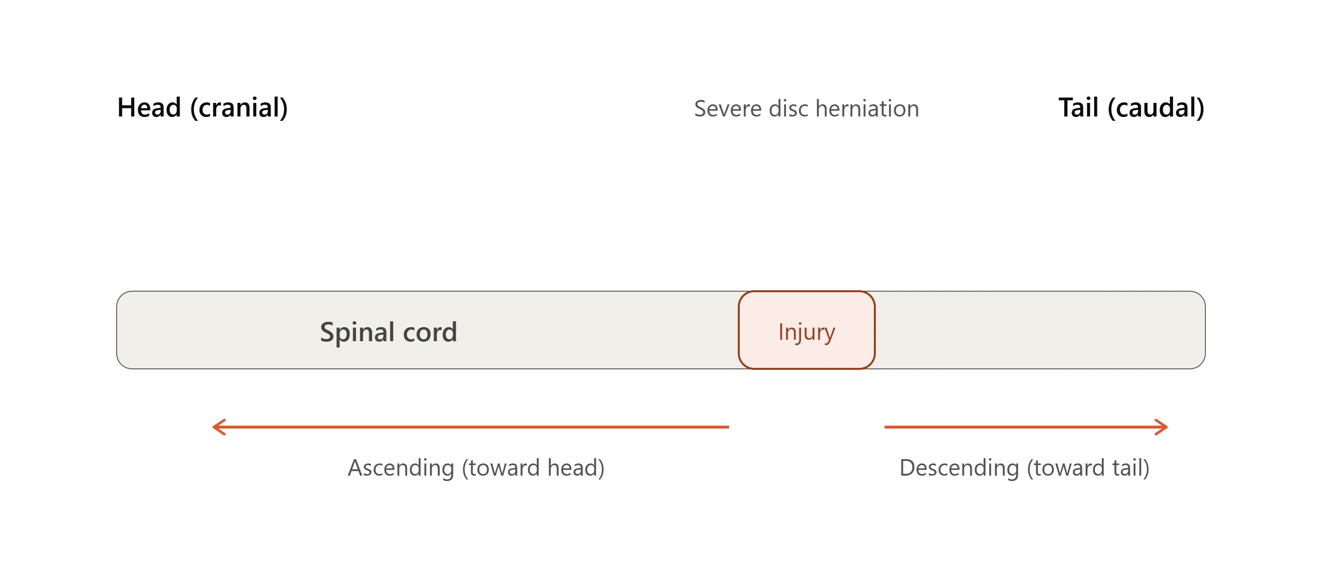
Myelomalacia is a condition in which spinal cord tissue softens and breaks down as nerve cells die. When discussing what myelomalacia is in dogs, the main concern is its progressive form. The damage begins at the site of a severe spinal injury and spreads along the spinal cord, sometimes in both directions.
- When it spreads toward the head, it is called ascending myelomalacia in dogs.
- When it spreads toward the tail, it is known as descending myelomalacia in dogs.
- In some dogs, the damage spreads in both directions, which is called ascending descending myelomalacia.
Progressive myelomalacia is usually fatal. A 2025 case-control study published in the Journal of Veterinary Internal Medicine described it as an uncommon but often fatal complication of a sudden, severe disc herniation in the middle or lower back.
Myelomalacia Cause and Pathophysiology
The most common cause of myelomalacia in dogs is a sudden disc herniation in the middle or lower back, known as Hansen Type I disc extrusion. In some cases, it develops after other severe forms of spinal cord injury in dogs, such as fibrocartilaginous embolism, which blocks blood flow to the spinal cord. Although myelomalacia usually affects the middle or lower back, a 2023 case report documented progressive myelomalacia after a neck disc extrusion.
Primary Injury and Secondary Injury
The primary injury occurs when the spinal cord is compressed and bruised by the extruded disc material. Secondary injury may be caused by ischemia, hemorrhage, free radical injury, and release of inflammatory enzymes. These changes injure nerve tissue and small blood vessels, allowing the damage to spread beyond the original injury site. However, the detailed mechanisms of spinal cord damage spread are still not well understood.
Risk Factors of Myelomalacia in Dogs
Earlier retrospective studies have identified several possible risk factors for progressive myelomalacia. However, these associations have not been consistently confirmed, and no single factor reliably predicts which dogs will develop the condition.
Risk factors reported across retrospective studies include:
- Absent deep pain perception with paraplegia at presentation
- Hyperacute onset of signs
- Younger age, with one study noting dogs under 6 years
- Disc extrusion in the lumbar intumescence, including L5 to L6
- French Bulldog breed
- Unneutered status, especially males (reported in 2025, but the study was likely underpowered)
In a 2025 case-control study of 244 dogs, researchers found some evidence of associations with disc extrusion involving the lumbar intumescence and French Bulldog breed, but the study lacked sufficient statistical power to confirm either association.
Age and the speed of onset were not significantly associated with Progressive Myelomalacia (PMM) in this study.
Signs and Symptoms
.png)
Clinical signs of progressive myelomalacia typically develop within days of the initial injury and may worsen rapidly.
Watch for these symptoms of myelomalacia in dogs:
- Loss of muscle tone and reflexes in the pelvic limbs, with a flaccid anus
- A cutaneous trunci cutoff that moves cranially over serial examinations
- Spinal pain or hyperesthesia that was not present before
- Hyperthermia and a new, unexplained decline in demeanor
- Hind limb weakness progressing to thoracic limb weakness
- Diaphragm paralysis and respiratory failure at the end stage
|
Finding |
What it suggests |
|
Cranial migration of cutaneous trunci cutoff |
Ascending spread of necrosis |
|
Loss of tail and anal tone, dilated anus |
Sacral segment involvement |
|
Reduced or absent thoracic limb reflexes |
Cervical intumescence involvement |
|
Intercostal and diaphragm weakness |
Imminent respiratory failure |
|
New spinal pain with fever |
Possible progressive myelomalacia |
One caution applies to the second row. Reduced reflexes right after injury can be spinal shock, so the trend across repeat exams matters more than a single finding.
Diagnosis
Veterinarians diagnose myelomalacia by observing how the dog's symptoms develop over time and reviewing imaging results, such as MRI scans. A confirmed diagnosis can only be made by examining spinal cord tissue under a microscope (histopathology).
- Neurologic examination: Record deep pain perception, reflexes, and the cutaneous trunci level at presentation, then repeat at set intervals.
- MRI: Okada and colleagues described MRI features associated with presumptive and confirmed progressive myelomalacia in 12 dogs, including extensive intramedullary signal change. Findings are supportive, not pathognomonic.
- Gradient echo sequences: A 2023 study of 82 paraplegic dogs without nociception found no significant difference in progression rates tied to this sequence, so interpret it cautiously.
- Cerebrospinal fluid analysis: It may flag progression before overt signs, but it is not a standalone test.
Differentials include spinal shock, ongoing compression, and severe nonprogressive cord injury.
Myelomalacia Treatment in Dogs and Surgical Options for Spinal Cord Injury in Dogs
There is no proven myelomalacia treatment in dogs that can stop spinal cord necrosis from progressing. Care focuses on supporting the dog and discussing humane options with the owner if the condition continues to progress.
Decompressive Surgery
Decompressive surgery is commonly considered for dogs with severe thoracolumbar intervertebral disc extrusion and absent deep pain perception, where appropriate.
Published evidence suggests that approximately 61% of surgically treated paraplegic dogs without pain perception recover ambulation, compared with 93% of dogs with preserved nociception.
These figures describe outcomes following IVDE-related spinal cord injury and should not be interpreted as recovery rates for dogs with established progressive myelomalacia.
Durotomy and Extended Hemilaminectomy
Some clinicians perform durotomy or extensive hemilaminectomy in dogs with presumptive progressive myelomalacia. A 2025 BMC Veterinary Research study compared two extensive hemilaminectomy and durotomy techniques in this population. Consider this an emerging area with limited evidence, not a recommended standard.
Supportive Care and Euthanasia
Supportive care covers analgesia, bladder management, thermoregulation, and ventilatory monitoring. Mechanical ventilation does not change the underlying disease. When progression is confirmed, humane euthanasia is the usual outcome.
Explore our Surgical Procedures resource section for more veterinary guidance and educational resources.
Prognosis, Life Expectancy, and Survival
Once progression is established, life expectancy is short, generally a matter of days. Survival with myelomalacia in dogs is rare, and it is more accurate to speak of survival without progression in the original Intervertebral Disc Extrusion (IVDE) population.
|
Data point |
Value |
Source |
|
Prevalence of Progressive Myelomalacia (PMM) in paraplegic dogs without deep pain perception |
10 to 17.5% |
BMC Vet Res, 2025 (summarizing earlier studies) |
|
PMM in paraplegic French Bulldogs without nociception |
33% |
BMC Vet Res, 2025 |
|
Recovery of ambulation after surgery, nociception absent |
61% overall |
Gomes et al., Front Vet Sci, 2024 |
|
Recovery of ambulation after surgery, nociception present |
93% |
Gomes et al., Front Vet Sci, 2024 |
When discussing progressive myelomalacia in dogs with owners, separate what is established (poor prognosis once progression is confirmed) from what is uncertain (individual risk at presentation).
Preventing Myelomalacia in Dogs
There is no proven method for preventing myelomalacia in dogs, and the 2025 JVIM study was designed to look for clinician-controlled factors that could lower risk. It did not identify any with confidence. Practical risk reduction is therefore upstream:
- Reduce Intervertebral Disc Extrusion (IVDE) risk in chondrodystrophic breeds with weight control and limiting repetitive jumping
- Refer promptly when a dog loses motor function
- Handle and transport dogs with suspected spinal injury carefully
- Reassess neurologic status at set intervals so early progression is not missed
FAQs
How fast does myelomalacia progress in dogs?
Signs generally emerge within days of the injury and can advance over hours to a few days, with the cutaneous trunci cutoff moving cranially. Exact timing varies between dogs.
Does myelomalacia in dogs occur after surgery?
Yes. Progression can follow decompressive surgery, which treats compression but does not stop necrosis that is already underway. Dogs with absent deep pain perception should be counseled on this before surgery.
How common is myelomalacia in dogs?
It is uncommon overall. Reported prevalence is 10 to 17.5% in paraplegic dogs without deep pain perception after IVDE, and one study found 33% in French Bulldogs.
How can myelomalacia in dogs be prevented?
No intervention is proven. Focus on lowering Intervertebral Disc Extrusion (IVDE) risk, early referral, and serial neurologic examination.
How do I tell progressive myelomalacia from spinal shock?
Spinal shock is usually stable or improves on repeat exams, while myelomalacia is progressive. Cranial migration of the cutaneous trunci cutoff, new spinal pain, fever, and MRI findings favor myelomalacia.
Conclusion
Myelomalacia in dogs is a rare but severe complication of acute spinal cord injury, concentrated in dogs with Grade 5 Intervertebral disc disease (IVDD). Early recognition through serial neurologic exams, appropriate imaging, and honest prognostic communication let veterinarians guide owners before a dog suffers unnecessarily. Be clear about where the evidence is solid and where it is still emerging.
Sources
- Castel A, et al. Risk factors associated with progressive myelomalacia in dogs with complete sensorimotor loss following intervertebral disc extrusion: a retrospective case control study. BMC Veterinary Research, 2019.
- Okada M, et al. Magnetic resonance imaging features and clinical signs associated with presumptive and confirmed progressive myelomalacia in dogs: 12 cases (1997 to 2008). JAVMA, 2010.
- Case control investigation of clinician determined variables with progressive myelomalacia after acute thoracolumbar disc extrusion in dogs. Journal of Veterinary Internal Medicine, 2025;39(4):e70180.
- Gomes SA, et al. Characterization of risk factors for early ambulation in paraplegic dogs with absent pain perception undergoing decompressive surgery for thoracolumbar intervertebral disk extrusions. Frontiers in Veterinary Science, 2024.
- MRI and progressive myelomalacia in 82 paraplegic dogs without nociception. Frontiers in Veterinary Science, 2023.
- Outcomes of non limited versus cranial limited extensive hemilaminectomy and durotomy in dogs with presumptive progressive myelomalacia. BMC Veterinary Research, 2025.
- Case report: progressive myelomalacia after cervical intervertebral disk extrusion. Frontiers in Veterinary Science, 2023.


