




Dog Paw Yeast Infection: Symptoms, Causes, Treatment, and Prevention
Key Takeaways
- A dog paw yeast infection is an overgrowth of Malassezia pachydermatis, a yeast that lives normally on canine skin, producing the inflammatory condition known as yeast dermatitis.
- Paw yeast overgrowth is almost always secondary. Allergic skin disease, endocrine disorders, and keratinisation defects are the most common underlying drivers.
- Cytology is the diagnostic standard. Lesion appearance alone cannot distinguish yeast dermatitis from bacterial pododermatitis, demodicosis, or neoplasia.
- The 2009 systematic review by Negre and colleagues found good evidence for one topical protocol: a shampoo containing 2% miconazole nitrate plus 2% chlorhexidine gluconate, applied twice weekly for three weeks.
- Recurrence signals an unaddressed primary disease. Repeated antifungal courses without a diagnostic workup will not resolve the case.
What Is a Dog Paw Yeast Infection?
A dog paw yeast infection is a fungal infection of the paw skin caused by overgrowth of commensal Malassezia yeast. The resulting inflammatory skin condition is termed Malassezia dermatitis, or yeast dermatitis.
Malassezia pachydermatis is a lipophilic yeast and a normal component of the canine skin mycobiota. It is present on healthy skin without causing disease. Pathology develops when population sizes increase, when the skin barrier is compromised, or when the patient mounts a hypersensitivity response to the organism.
Pododermatitis describes inflammation of the paw skin from any cause. Yeast dermatitis is one cause among several, which is why the two terms are not interchangeable and why a cytological diagnosis matters.
The WAVD Clinical Consensus Guidelines note that Malassezia dermatitis has moved from a condition whose existence was once disputed to a routine diagnosis in general practice.
What Does a Dog Paw Yeast Infection Look Like?
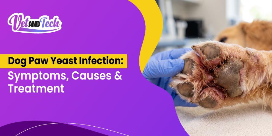
Paw yeast dermatitis presents with pruritus, erythema, malodour, and progressive skin changes. Owners typically report the behavioural signs before the dermatological changes are recognised.
Clinical signs commonly seen:
- Licking and chewing at the paws, particularly the interdigital spaces. The behaviour is self-perpetuating: saliva adds moisture, and repeated trauma degrades the barrier further.
- Erythema and irritation between the toes and around the paw pads.
- Reddish-brown discoloration of the fur. Important distinction: this staining is caused by saliva porphyrins from repeated licking, not by the yeast itself.
- Brown discharge at the nail base, sometimes extending onto the nail plate.
- Malodour, often described by owners as musty or corn chip-like.
- Flaky, scaly, greasy, or thickened skin, progressing to lichenification and hyperpigmentation in chronic cases.
- Alopecia around the digits, secondary to self-trauma rather than the infection.
- Interdigital swelling as inflammation becomes more pronounced.
- Lameness or resentment of paw handling when inflammation is marked.
- Fissures, erosions, and ulceration in long-standing cases, with secondary bacterial infection once the barrier is breached.
Why Do Dog's Paws Smell Like Corn Chips?
The corn chip odour comes from normal skin flora, including commensal bacteria and yeast, metabolising on the skin surface. On its own it is frequently a normal finding.
Odour becomes clinically relevant when it accompanies erythema, pruritus, staining, or discharge. An owner reporting "Frito feet" with no other changes usually does not have a patient with yeast dermatitis. This distinction is worth making explicitly during the consultation, because it is a common reason for unnecessary owner concern and unnecessary treatment.
These Signs Are Not Specific to Yeast
None of the above findings confirm Malassezia dermatitis. Flea allergy dermatitis, atopic dermatitis, adverse food reactions, bacterial pododermatitis, demodicosis, foreign body reactions, and less commonly skin cancer can all produce overlapping presentations.
|
Differential |
Suggestive features |
Confirmatory approach |
|
Malassezia dermatitis |
Malodour, greasy or lichenified skin, salivary staining |
Cytology showing budding yeast |
|
Bacterial pododermatitis |
Papules, pustules, purulent discharge, epidermal collarettes |
Cytology showing cocci; culture if indicated |
|
Atopic dermatitis |
Bilateral distribution, seasonality, concurrent otitis and facial or axillary involvement |
Clinical criteria plus exclusion of other causes |
|
Flea allergy dermatitis |
Dorsal lumbar involvement, flea evidence |
Flea combing, response to rigorous flea control |
|
Demodicosis |
Alopecia, comedones, follicular casts |
Deep skin scrapings, trichogram |
|
Foreign body reaction |
Unilateral, single interdigital site, draining tract |
Imaging, exploration |
|
Neoplasia |
Solitary persistent mass, non-responsive lesion |
Biopsy and histopathology |
Unilateral single-digit disease that does not respond to appropriate antifungal therapy deserves particular scrutiny. Foreign body and neoplastic causes are underdiagnosed in this presentation.
What Causes a Yeast Infection on a Dog's Paw?
Paw yeast dermatitis develops when Malassezia pachydermatis proliferates beyond commensal levels. Three conditions drive that proliferation: moist skin, poor airflow, and a compromised barrier.
Moisture. Interdigital spaces retain water from wet grass, rain, snow, swimming, bathing, and saliva. Warm, moist skin favours Malassezia growth.
Restricted airflow and skin folds. Yeast overgrowth concentrates in anatomical sites with limited ventilation. The classic examples are skin folds, the ear canal, and the interdigital spaces, which function much like skin folds. Dense interdigital hair worsens moisture retention.
Barrier compromise. This is the decisive factor. Yeast is present on healthy paws without consequence. Disease requires something to disrupt the barrier or the local immune response first, which is why treating the yeast without identifying that primary problem produces temporary improvement followed by relapse.
The relationship is more complex than simple overgrowth. In dogs with allergic skin disease, Malassezia can act as an allergen and provoke a hypersensitivity response, amplifying pruritus and inflammation beyond what organism numbers alone would explain.
Which Dogs Are Most at Risk?
Any dog can develop paw yeast dermatitis, but several groups are overrepresented.
Dogs with allergic skin disease. The largest risk group by a wide margin. Breeds predisposed to atopy, including Golden Retrievers, Labrador Retrievers, Boxers, Shih Tzus, Dalmatians, and Staffordshire Bull Terriers, are commonly reported with pododermatitis and secondary yeast overgrowth.
Breeds with prominent skin folds or dense coats. Shar Pei, English Bulldog, and French Bulldog patients retain moisture in folds and interdigital spaces. Heavily coated feet, as in Cocker Spaniels and Poodles, hold moisture after exposure to wet ground.
Breeds commonly reported as predisposed to Malassezia overgrowth. West Highland White Terriers, Basset Hounds, Cocker Spaniels, Dachshunds, Shetland Sheepdogs, Lhasa Apsos, Maltese, and Poodles appear repeatedly in the dermatology literature. West Highland White Terriers in particular are strongly associated with Malassezia dermatitis and epidermal dysplasia.
Senior patients. Two contributing factors. Immune competence declines with age, and osteoarthritis or reduced mobility limits self-grooming, allowing debris and moisture to accumulate between the toes.
Dogs with high moisture exposure. Working dogs, swimmers, dogs walked on wet ground, and dogs bathed frequently.
What Facilitates Yeast Overgrowth?
Yeast overgrowth is a secondary problem in the great majority of cases. The primary conditions most often responsible:
- Environmental allergies. Pollen, dust mites, and mould producing atopic dermatitis.
- Flea allergy dermatitis. Hypersensitivity to flea saliva driving inflammation and pruritus, including around the paws.
- Adverse food reactions. Food sensitivity contributing to recurrent pruritus and cutaneous inflammation in a subset of patients.
- Endocrine disorders. Hypothyroidism and hyperadrenocorticism produce cutaneous changes that predispose to secondary Malassezia overgrowth.
- Keratinisation disorders. Zinc-responsive dermatosis, vitamin A-responsive dermatosis, and primary seborrhoea. These are underconsidered and worth including in the differential list for refractory cases.
- Bacterial pododermatitis. Frequently concurrent rather than alternative.
- Trapped moisture. A contributing factor rather than a primary disease, but sufficient to sustain overgrowth once the barrier is compromised.
- Local trauma or foreign bodies. Grass awns, wounds, and puncture injuries breaching the barrier directly.
- Neoplasia. Uncommon, but skin cancer can compromise local tissue and should remain on the list for solitary non-responsive lesions.
How Do Veterinarians Diagnose Paw Yeast Infection?
Cytology is the diagnostic standard. Lesion appearance alone cannot confirm Malassezia dermatitis, and treating on appearance alone risks missing bacterial, parasitic, or neoplastic disease.
Sampling from paw skin can be performed by acetate tape preparation, impression smear, or another appropriate technique matched to the site. Acetate tape is generally the most practical for interdigital skin. Samples are stained and examined microscopically for the characteristic oval, peanut-shaped, or footprint-like budding yeast.
A positive cytology does not establish causation. Two points matter here. First, Malassezia is a normal skin resident, so its presence must be interpreted against organism numbers, clinical signs, and history. Second, in allergic patients the organism can act as an allergen, meaning clinical severity may exceed what organism counts suggest.
Cytology should also be assessed for cocci. Concurrent bacterial infection is common and changes the treatment plan.
Further diagnostics are directed by history and physical examination, and may include deep skin scrapings for Demodex, trichogram, dermatophyte testing, endocrine testing, elimination diet trial, or biopsy for persistent or atypical lesions. Note that allergy testing is used to formulate allergen-specific immunotherapy rather than to diagnose atopic dermatitis, which remains a clinical diagnosis.
How Do Veterinarians Treat Yeast Infections in Dog Paws?
Treatment of paw yeast dermatitis focuses on controlling the Malassezia overgrowth and addressing any underlying skin diseases that may be contributing to recurrence.
Depending on the extent and location of the disease, veterinarians may use topical therapy alone or combine topical treatment with systemic antifungal therapy.
When there is little or no improvement with antifungal therapy, other potential causes should be evaluated. Bacterial infection, allergies, foreign bodies, and neoplasia should be considered in the evaluation of persistent or unusual lesions.
Topical Treatments for Yeast Dermatitis in Dogs
Topical antifungal therapy is often used for localized disease. Medicated shampoos, wipes, rinses, and other topical products may be selected based on the location and extent of the lesions. Topical products can also be used alongside systemic medication when appropriate.
A systematic review found good evidence for a shampoo containing 2% miconazole nitrate and 2% chlorhexidine gluconate for treating canine Malassezia dermatitis. In the studies reviewed, the shampoo was applied twice weekly for three weeks.
Medicated wipes and leave-on products may also be used for localized paw lesions or between bathing treatments, depending on the product and the individual case. These products allow topical treatment to reach the spaces between the toes.
Oral Antifungal Medications for Yeast Dermatitis in Dogs
For dogs with generalized disease, difficult-to-manage cases, or cases that do not respond adequately to topical treatment, veterinarians may consider systemic antifungal therapy such as itraconazole or ketoconazole. Itraconazole and ketoconazole are systemic antifungal options used for canine Malassezia dermatitis.
Treatment and dose, monitoring requirements, patient-specific factors, and the appropriate drug will vary by individual dog; veterinary references should be consulted, and patient-specific factors should be considered prior to treatment.
Potential Side Effects of Azole Antifungals
Ketoconazole and itraconazole are metabolized in the liver, with possible interactions with other medications via the cytochrome P450 system. Liver enzymes and other laboratory parameters may be monitored by the veterinarian for dogs on systemic azole therapy depending on the drug used, duration of therapy, and individual patient factors.
Why Topical Treatment Sometimes Fails
Malassezia can form biofilm, an extracellular matrix that promotes adherence to the skin surface and reduces the effective penetration of topical antimicrobials. Biofilm formation has been documented in co-cultured M. pachydermatis, and it is one explanation for apparent topical failure in a patient with a correct diagnosis.
Before escalating therapy, three other explanations deserve checking:
- Owner compliance and technique. Insufficient contact time, inadequate application between the toes, or missed applications.
- Concurrent bacterial infection not covered by the chosen product.
- An unaddressed primary disease driving continued inflammation regardless of organism control.
Persistent or unusual lesions that do not respond to appropriate therapy warrant reassessment for bacterial infection, allergic disease, foreign body, and neoplasia.
Managing Pruritus and Inflammation Alongside Antifungal Therapy
Controlling the organism does not immediately resolve the itch, and continued licking sustains the moisture and trauma that permitted overgrowth in the first place.
Where allergic disease is driving the presentation, anti-inflammatory or anti-pruritic medication may be used alongside antifungal therapy to interrupt that cycle. Options include glucocorticoids for short-term control, and targeted anti-pruritic therapies for longer-term management of atopic dermatitis. Selection, duration, and monitoring should follow current atopic dermatitis treatment guidance and the individual patient's history.
Barrier support measures, including essential fatty acid supplementation, are commonly used as part of long-term management in allergic patients.
How Long Does Treatment Take?
The best-supported topical protocol runs three weeks. Uncomplicated localised cases often improve within that window.
Recurrent and complicated cases take considerably longer, and expectations should be set with the owner at the first consultation. Where an underlying allergic or endocrine disease is present, the realistic goal is long-term control with maintenance topical therapy at intervals determined by clinical response, not a single curative course.
Recheck appointments allow confirmation of cytological resolution, assessment of treatment tolerance, and reassessment when response is inadequate. Clinical improvement without cytological resolution is a common reason for early relapse.
Home Care and What to Avoid
Owner-managed measures support veterinary treatment. They do not substitute for diagnosis, and owners should confirm any home measure with the attending veterinarian before starting.
Supportive and generally low-risk:
- Drying the paws thoroughly after wet walks, swimming, or bathing, with attention to the interdigital spaces.
- Clipping excess interdigital hair where it retains moisture.
- Veterinary-recommended antifungal wipes for maintenance between baths.
- A gentle, unscented soap or antiseptic paw soak to remove surface debris, taking care that the patient does not drink the solution.
Requires veterinary discussion first:
- Apple cider vinegar. Frequently recommended online and worth cautioning against unsupervised use. It can sting and cause discomfort on eroded or fissured skin, and excessive drying is counterproductive when the barrier is already compromised.
- Tea tree oil products. Concentration matters. Higher concentrations carry toxicity risk in dogs.
- Dietary change. A meaningful intervention where adverse food reaction is suspected, but an over-the-counter novel protein diet is not equivalent to a diagnostic elimination diet trial. Over-the-counter diets are unsuitable for a true food trial because of cross-contamination.
- Probiotics and supplements. May have a role in broader skin health, but should not delay diagnosis of the primary disease.
Not appropriate: human antifungal preparations used without veterinary direction, and repeat courses of leftover prescription medication.
Frequently Asked Questions
Can a dog paw yeast infection go away on its own?
Uncommonly. Because the yeast is a normal skin resident and the paws continue to trap moisture, established yeast dermatitis usually persists or worsens without treatment. Self-trauma from licking sustains the barrier damage that permitted overgrowth.
How long does it take to clear a paw yeast infection?
The best-supported topical protocol runs three weeks. Uncomplicated localised cases often improve within that period. Recurrent cases with an unmanaged underlying allergy require long-term control rather than a single course.
Is a dog paw yeast infection contagious to humans or other dogs?
Malassezia pachydermatis is a normal canine skin commensal, and transmission to healthy household dogs or people is not a routine concern. Rare human infections have been reported in immunocompromised patients in healthcare settings, which is a distinct scenario from ordinary pet ownership.
Do dogs need a diet change for yeast infections?
Only where adverse food reaction is contributing. That requires a properly conducted elimination diet trial rather than an over-the-counter novel protein food, which is unsuitable for diagnostic purposes because of cross-contamination.
Can dogs take fluconazole for yeast infections?
It is sometimes prescribed, but it is generally considered less effective against Malassezia than itraconazole or ketoconazole. It is not a first-line equivalent, and any systemic azole requires drug interaction review and appropriate monitoring.
What is the difference between a bacterial and a yeast paw infection?
Cytology distinguishes them: budding yeast versus cocci. Clinically, bacterial pododermatitis more often shows papules, pustules, and purulent discharge, while yeast dermatitis more often shows greasy or lichenified skin with malodour. The two occur together frequently, which is a further reason to sample rather than assume.
Conclusion
Yeast infections can make dogs uncomfortable, but treating the yeast is only part of the picture. Because Malassezia dermatitis is often associated with underlying allergic or other skin diseases, recurring cases may require investigation into what is driving the inflammation. Depending on the dog and the extent of the disease, antifungal shampoos, other topical treatments, and systemic medications may all have a place. Persistent licking, odor, redness, or recurring paw problems warrant a veterinary examination. Cytology can help identify Malassezia, while the dog's history and additional diagnostic tests can help identify factors contributing to recurrence.
Learn More About Veterinary Dermatology
Recurrent paw yeast infections have varied underlying causes, and staying current on dermatology helps when these cases keep returning.
Vet and Tech offers free, RACE-approved CE sessions covering skin disease, dermatological diagnostics, allergy management, and treatment approaches.
Browse the webinar library to find upcoming and on-demand sessions for your practice.
References
- Bond R, Morris DO, Guillot J, Bensignor EJ, Robson D, Mason KV, Kano R, Hill PB. Biology, diagnosis and treatment of Malassezia dermatitis in dogs and cats: Clinical Consensus Guidelines of the World Association for Veterinary Dermatology. Veterinary Dermatology. 2020;31(1):28-74. doi:10.1111/vde.12809 (open access)
- Negre A, Bensignor E, Guillot J. Evidence-based veterinary dermatology: a systematic review of interventions for Malassezia dermatitis in dogs. Veterinary Dermatology. 2009;20(1):1-12. doi:10.1111/j.1365-3164.2008.00721.x
- Guillot J, Bensignor E, Jankowski F, et al. Comparative efficacies of oral ketoconazole and terbinafine for reducing Malassezia population sizes on the skin of Basset Hounds. Veterinary Dermatology. 2003;14(3):153-157.

%20VAT%20Blog%20-%20Remote%20Veterinary%20Jobs%20Roles%2C%20Pay%20%26%20How%20to%20Find%20Them-thumbnail-1790256372.webp&w=3840&q=75)


