


Ear Surgery
Eye Surgery
Orthopedic Surgery
Soft Tissue Surgery
- Caudectomy Treatment
- Cholecystectomy (Gallbladder Removal)
- Brachycephalic Obstructive Airway Surgery
- Cystotomy Surgical Procedure
- Diaphragmatic Hernia Repair
- Gastric Dilatation-Volvulus (GDV)
- Treat recessed vulva with Episioplasty
- GI Foreign Body Removal
- Mass / Tumor Removal in Pets
- Surgical procedure of cystotomy and Urethrostomy
- Liver Shunt - Constrictor Ring Application Procedure
- GDV Correction with Gastropexy Surgical Procedure
- Mastectomy surgical procedure
- Nephrectomy surgical procedure
- Partial Thyroidectomy Surgical Procedure
- Parathyroidectomy surgical procedure
- Everything You Must Know About The Perineal Hernia Surgery
- The Perineal Hernia Surgery With Diverticulum In Dogs
- Prepuceplasty: A Detailed Insights Into Treatment and Complications
- Pyometra Surgery For The Female Dogs
- A Comprehensive Analysis Of Sialocele Surgical Procedure
- Urethrostomy – Prevention of a life-threatening condition
- Anal Gland Removal (Unilateral or Bilateral)
- A Reliable Treatment For Chronic Constipation
- Wound Closure with Skin Graft
- Wound Closure With Stretching Device In Animals
Bulla Osteotomy_Procedural Details, Complications, Cost, & More
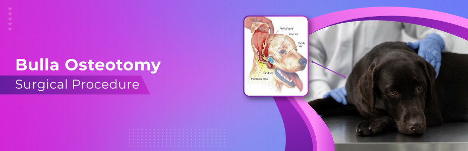
Chronic ear infections can be a nightmare for the pet's owner as there is a high chance that the pet will become deaf. In addition, the continual growth of the ear infection can make its treatment even more complicated. However, due to technological advancement in the medical industry, veterinarians can deal with almost all inflectional diseases. Indeed, the ear canal is narrow, and it is impossible to clean your pet's infected ears at home. Therefore, the final option is to go for a surgical treatment like bulla osteotomy.
In most cases, it's all about the diseased tissues. The surgery includes the removal of the entire ear canal; the middle ear goes through draining to remove the infected material, and then the ear is closed. Undoubtedly, it is painful and involves plenty of complications that we will discuss below. Most pet owners must be aware of this structure (bulla).
Why Is Bulla Osteotomy?
Bulla osteotomy surgical procedure is used primarily in veterinary medicine to treat middle ear diseases, particularly in dogs. The term "bulb" refers to the tympanic bulb, the hollow bony structure at the base of the ear that contains the structures of the middle and inner ear. "Osteotomy" is a medical term for cutting or removing part of a bone.
A tympanic osteotomy involves surgically opening or removing part of the cochlea to allow direct access to the middle ear. In this way, the veterinarian can resolve problems such as chronic ear infections and the buildup of foreign bodies that cause discomfort or pain in the animal.
Bulla osteotomy dog can be performed using various surgical techniques, such as laterally (sideways), ventrally (downward), or a combination of these, depending on the type of problem and the animal's anatomy.
Indications For Bulla Osteotomy
Bulla osteotomy surgery procedure is generally performed when more conservative treatments for middle ear problems, such as antibiotics or topical medications, have failed. Indications for bulla osteotomy include:
- Chronic Otitis Media: This is a persistent or recurrent inflammation or infection of the middle ear. Chronic otitis media can be very uncomfortable for the animal and, if left untreated, can lead to more severe problems, including deafness.
- Tympanic Polyps: These benign tumors can appear in the middle ear and cause discomfort or affect normal hearing.
- Foreign Bodies: Foreign bodies can sometimes become lodged in the middle ear. This most often occurs in animals working outdoors or in the workplace.
- Neoplasia: Neoplasia is an abnormal tissue growth that can be benign or malignant. Sometimes, middle ear tumors must be removed by ventral bulla osteotomy.
- Middle Ear Effusions: This condition is characterized by an accumulation of fluid in the middle ear, often due to infection or inflammation.
Before performing an ear bulla osteotomy, diagnostic images such as X-rays, CT scans, or MRIs can be used to assess the extent of the problem and plan the surgical approach.
Results Of The Surgery
In many cases, the surgical results of bulla osteotomy cat or dog are miraculous. The patients feel more energetic and do not think of any headaches. There would not be any more lousy odor or pain to the patient. Even the ears will not require frequent cleaning. Above all, the surgical procedure requires modern technical skills, and some veterinarians feel uncomfortable performing this surgery. Thus, do discuss this in detail with your veterinarian so they can recommend the best specialist for your pet.
Pre-Operative Care For Bulla Osteotomy Surgical Procedure
Preparation for a bulla osteotomy begins with a thorough physical examination and review of the animal's medical history. Next, diagnostic tests, including blood and urine analyses, are performed to assess the animal's general health and suitability for anesthesia. In addition, imaging studies, such as X-rays, CT scans, or MRIs, are used to assess the exact condition of the ear and its structures, helping to plan the surgical procedure.
Before surgery, treating any ongoing ear infections with appropriate antibiotics, determined by culture and sensitivity tests, is essential. If the patient takes medication, the veterinarian will advise them on handling it before the operation. In addition, it may be necessary to deprive the patient of food and water for some time before anesthesia, depending on the veterinarian's instructions.
Intraoperative Care For Bulla Osteotomies
The patient's vital signs, including heart rate, blood pressure, respiratory rate, oxygen saturation, and temperature, are carefully monitored during the procedure. Anesthesia is administered to ensure the patient does not suffer or remain motionless during the process. Anesthesia during surgery is administered with a combination of local and systemic analgesics.
The surgical site is prepared by trimming the fur and cleaning the skin to reduce the risk of infection. Next, the surgeon uses specialized tools to make an incision and gain access to the tympanic pouch. The affected tissue or foreign body is removed, and any necessary repair or cleaning of the ear structures is performed.
Post-Operative Care For Bulla Osteotomy
Post-operative care is essential for recovering and healing after ear surgery. Pain management remains a priority, and analgesics are administered if necessary. The veterinarian may also prescribe antibiotics to prevent infection and anti-inflammatories to reduce swelling.
The surgical area should be kept clean and dry, and regular post-operative examinations should be carried out to monitor the healing process and remove sutures if necessary. A cone or electronic collar can be used to prevent the animal from scratching or shaking its head, which could hinder the healing process.
During bulla osteotomy recovery, your pet's activity should be limited according to the veterinarian's instructions, which often involves avoiding baths and exercise.
Any sudden behavior change, signs of pain, loss of appetite, or unusual discharge from the ear should be reported to the veterinarian immediately. A hearing test may also be carried out after the operation to detect any changes in the animal's hearing function.
Some animals may benefit from rehabilitation therapy after surgery to improve balance and motor function, as the operation can sometimes affect the vestibular apparatus, which helps control balance.
Rehabilitation After Bulbar Osteotomy
Bulla osteotomy recovery depends on each animal's condition, age, and general state of health. Most animals are generally better in the days to a week following the procedure. However, the complete healing process may take several weeks.
During this period, it is essential to follow the veterinarian's instructions regarding wound care, medication, feeding, and restriction of movement. In addition, the veterinarian should be consulted regularly to monitor the healing process, remove sutures if non-absorbable sutures have been used, and assess the healing progress.
Any complications, such as infection or recurrence of the original problem, should be monitored. In some cases, the nature of the procedure and its proximity to inner ear structures may result in temporary or permanent changes to hearing or balance. In addition, some animals may benefit from rehabilitation therapy to improve balance and motor skills.
Possible Complications
- The bulla is at the ear canal's bottom and is close to other essential ear structures. For instance, the systems are prone to damage throughout the surgical procedure. Even inflammation can occur in the healing process.
- Removing the tremendous auricular vasculature is another drawback of the bulla osteotomy procedure. For instance, the tissue of the great auricular vasculature dies along with the ear flaps. Therefore, the trimming of this portion is crucial as well. Other than that, there would not be any blood supply in the tremendous auricular vasculature.
- The retro glenoid vein can sometimes burst up, and the bleeding does not stop. Therefore, the visibility of the incision decreases. The retro glenoid vein is just beneath the bulla. Again, though, the bleeding reduces the procedure's efficacy.
- Sometimes, the swelling in the throat makes breathing difficult for the patient.
- There are 5 to 10 % cases of bulla osteotomy where the patient goes through chronic drainage due to the incision. Further, the second surgery is held to repair the issues. For example, the residual cells leftover in bulla and other fluids are drained from the eustachian tube.
- The facial nerves run near the ear's base. Also, it will control facial expressions. Unfortunately, facial paralysis is not expected after an ear disease. It means that the pet will have its jaw slacked on one side. As a result, the eyes would not be able to blink normally. After some time, the eyes will start retracting to assist in tear lubrication. Thus, the blinking loss does not cause any damage to the patient's eyes. Nonetheless, it is a common complication of total ear canal ablation (TECA), but sometimes it can be permanent (chances are 10 to 15 %).
Other Complications
Hearing can be limited after the surgical procedure. The hearing ability reduces due to the chronic ear infection. Thus, the hearing disruption should be considered because the eardrum is removed. Some of the patients still hear the sounds typically after TECA.
Another complication includes cholesteatoma, and it is crucial to remove otherwise, there will be continuing drainage from the affected area.
More than that, the patient will be suffering from oral pain while chewing or opening the mouth. Hence, a CT scan is vital for knowing the patient's condition and planning the surgical procedure appropriately.
Bulla Osteotomy Cost
The cost can vary considerably and depends on several factors, including the
- The complexity of the procedure
- Geographical location
- The veterinary clinic performing the procedure
Other factors likely to influence costs are
- Pre-operative diagnosis (blood tests, imaging)
- Anesthesia
- The procedure itself
- Medication and post-operative care
- Follow-up examinations.
In the United States, the cost of surgery can vary from $1,000 to $3,000 or more. Therefore, discussing potential costs with your veterinarian before surgery is essential so you can understand what to expect and plan financially for your pet's care.
Remember that the cost of veterinary services can change and increase over time. It is, therefore, advisable to consult your local veterinarian to obtain as accurate and up-to-date a cost estimate as possible. In addition, some pet insurance policies may cover all or part of the cost of procedures such as osteotomies or bone marrow biopsies.
Final Thoughts!
Bulla osteotomy is a delicate procedure that involves the removal of the ear canal as well as a tympanic bulla and leaves only the pinna (middle ear). As a result, the patient will have hearing loss no matter how successful the surgery goes. Mostly, the pets recover quickly after the surgery, but post-operative care is also necessary. The pet should rest for at least two weeks to let the wound heal properly. Overall, the prognosis of the surgery is reliable, but some complications are likely to occur.